
EMEGS (Qt version) offers an occular correction procedure based on
hierarchichal mutiple linear regression: the vertical (VEOG), the
horizontal (HEOG) and the radial electrooculogram (REOG) are used as
independant variables in a multiple linear regression for each EEG
channel. The corresponding beta weights (slopes) are tested for
statistical significance and non-significant channels are removed from
the model until only significant channels remain.
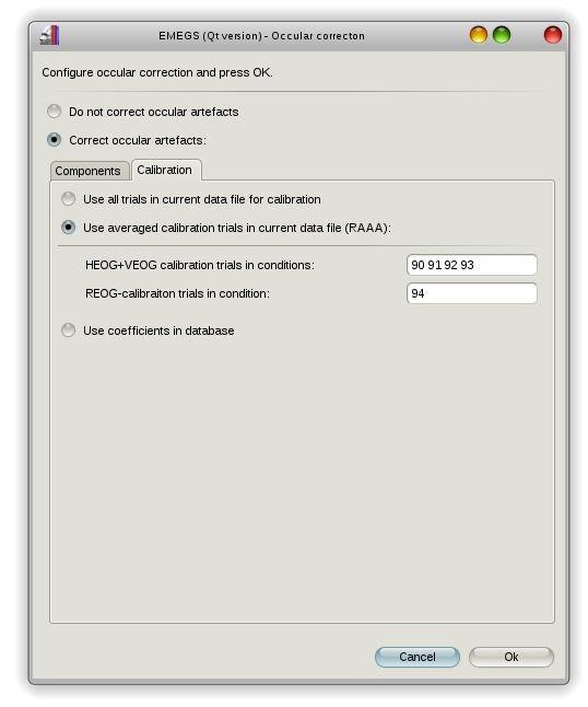
You can either use all your experimental data to calulate the beta
weights or use the revised artefact-aligned-average procedure (RAAA) as
suggested by Croft & Barry (2000). This procedure requires data
from a specific calibration sequence and estimates VEOG/HEOG and REOG
betas separately, from saccade and blink data respectively.
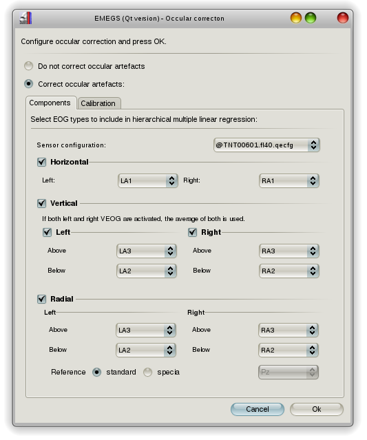
EMEGS (Qt version) will prompt you to configure the occular
correction when you press the "Resume analysis"-button after the data
segmentation step. Here you can select the sensors for VEOG, HEOG and
REOG. During previous steps, EMEGS (Qt versions) usually creates an
individual sensor configuration file next to the data file (to account
for instance for dropped channels when processing BDF files, that can
contain non-EEG channels). This file is selected by default and an @
-sign is put in front of the filename to indicate, that ist is a
dynamically created individual file.
Sensor configuration can contain information for occular
corrections, namely the keywords HEOG_L, HEOG_R,
LVEOG_B,
LVEOG_T, RVEOG_B,
RVEOG_T and
REOGRef in the "Occular" column, indicating the left HEOG
sensor, right HEOG sensor, left-bottom VEOG sensor, left-top VEOG
sensor, right-bottom VEOG sensor, right-top VEOG sensor and REOG
reference sensor respectively. These channels will be selected
automatically, if a sensor configuration file (or an adopted version of
it) is selected. An example of a configuration file is shown below:
Type Name
InRawFile
RefWeight Occular
Theta
Phi Rho
EEG Fp1 Yes
0
No
-1.6057 -1.25664 0.09
EEG AF7
Yes
0
No
-1.6057 -0.942478 0.09
EEG AF3
Yes
0
No
-1.29154 -1.13446 0.09
EEG F1
Yes
0
No
-0.872665 -1.18682 0.09
... ...
...
...
...
...
... ...
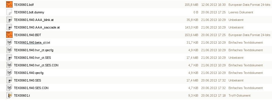
After the occular correction, the corrected trials are displayed,
and a couple of new files have appeared in your data folder, most
importantly the corrected SES-file, the name of which is constructed
depending on your occular correction configuration (hvr
for HEOG, VEOG and REOG, _ct
for calibrated with calibration trials, or
_at for calibrated with all trials). A
text-file containing the regression betas is also created, which looks
like this:
...
Sensor P3:
-----------------------------------------------------------------------------------------------------
Component Beta
Std.dev.
T
P
Step
Intercept -9.66859498e-12
1.64139629e-01 -5.89046962e-11
1.0000000 2
VEOG
1.17232219e-01 3.85906319e-03
3.03784139e+01 0.0000000* 2
HEOG
1.66331788e-03 1.29132049e-03
1.28807518e+00 0.1987550 1
REOG
-3.53498120e-01 3.63414858e-03
-9.72712348e+01 0.0000000* 1
...
If the RAAA-procedure was used, two additional files are created
(*AAA_saccade.at & *AAA_blink.at) containing the artefact-aligned
averages.
Croft, R.J., Barry, R.J. (2000). Removal of ocular artifact from the
EEG: a review. Clinical neurophysiology, 30(1), 5-19.